Category: Animals

  • New evidence suggests spinosaurus spent most of its life underwater

    New evidence suggests spinosaurus spent most of its life underwater

    For millions of years, evolution helped animals make the transition from life in the ocean to life on land. But the journey did not always move in one direction. Many species eventually returned to the water, developing new adaptations that allowed them to thrive in aquatic environments.

    Dozens of major groups of mammals and reptiles made this remarkable evolutionary reversal. Now, a new study from Yale University is shedding light on when and how these transitions occurred, as well as which ancient animals became fully aquatic and which continued to depend on land.

    The findings were published in the journal Current Biology.

    “These secondarily aquatic groups adapted in strikingly similar ways to their new aquatic home — evolving flippers and a suite of other features that made them better swimmers,” said lead author Caleb Gordon, who completed his doctorate at Yale’s Graduate School of Arts and Sciences (GSAS) earlier this year and is now a postdoctoral researcher at the Florida Museum of Natural History. “As a result, they’ve become textbook examples of convergent evolution, which can tell us a lot about the processes driving and constraining adaptive change in response to similar environmental cues.”

    Using Ancient Fossils and AI to Reconstruct Evolution

    To investigate these evolutionary changes, the researchers examined hundreds of fossil and modern specimens housed at the Yale Peabody Museum and dozens of other institutions worldwide. They collected extensive new data, including measurements (more than 11,000), photographs, and CT scans.

    The team combined traditional paleontological techniques with machine learning models that account for evolutionary relationships. They also incorporated a statistical approach originally used by naval forces during World War II. Together, these methods offered a new way to investigate one of the most remarkable transitions in vertebrate evolution.

    “Reconstructing the lives of extinct life forms in a scientifically rigorous way, as opposed to just telling stories, is a precise and delicate undertaking,” said Bhart-Anjan Bhullar, senior author of the study. Bhullar is an associate professor of Earth and planetary sciences in Yale’s Faculty of Arts and Sciences (FAS) and an associate curator of vertebrate paleontology and vertebrate zoology at the Peabody Museum.

    “It requires a careful interweaving of data from modern organisms, of which our understanding is necessarily far deeper, and knowledge of these living organisms’ genealogy relative to fossil forms,” he said. “Caleb’s work accomplishes all of this and more, and on a massive evolutionary scale.”

    For decades, scientists have disagreed about when particular groups of prehistoric animals became fully adapted to life underwater. The fossil record often provides incomplete or conflicting clues, making it difficult to determine whether an extinct species primarily lived on land, in water, or in both environments.

    Some skeletal features may indicate an aquatic lifestyle, while others suggest that the same animal remained capable of moving and feeding on land. These contradictions have led researchers to different conclusions about how ancient species behaved.

    “In these cases, paleontologists are often stuck, as different lines of evidence disagree about what the ancient animal was like,” Gordon said.

    To address this uncertainty, Gordon developed a method using relatively simple but effective machine learning models trained on living animals. By identifying relationships between skeletal proportions and known lifestyles, the models can estimate how extinct species used aquatic environments. They can also predict soft tissue adaptations, including swimming structures that rarely survive in fossils.

    Spinosaurus May Have Spent Most of Its Life Underwater

    One of the most striking findings concerns Spinosaurus, a giant prehistoric dinosaur whose relationship with water has generated considerable controversy.

    Spinosaurus lived approximately 113 million to 94 million years ago in what is now northern Africa. Although scientists generally agree that it was associated with aquatic environments, exactly how it moved, hunted, and spent its time remains a subject of debate.

    Some evidence points to an animal that frequently submerged itself and pursued prey underwater, much like seals or penguins. Other findings suggest a lifestyle closer to that of a modern heron, with Spinosaurus walking and searching for food along shorelines and in shallow water.

    The Yale team’s analysis provides new support for the more aquatic interpretation.

    “Our results shed new light on how much time Spinosaurus spent submerged, which could support the underwater hunting view,” Gordon said. “We confidently recovered highly aquatic habits for Spinosaurus, indicating that it spent the vast majority of its time submerged in the water.”

    Although the findings do not settle every question about how Spinosaurus hunted, they strengthen the case that this enormous predator spent much more of its life underwater than on land.

    Ancient Marine Reptiles That Never Fully Left Land

    The researchers reached a different conclusion about mesosaurs, small marine reptiles that lived between 290 million and 274 million years ago in regions now forming parts of South Africa and eastern South America.

    Mesosaurs have long interested paleontologists because they gave birth to live young, a characteristic some researchers have interpreted as evidence of a fully aquatic existence. However, the new analysis suggests that their limbs were not adapted for a life spent entirely in water.

    Instead, their skeletal proportions indicate that mesosaurs regularly moved between aquatic and terrestrial environments.

    “They spent a lot of time on land, like a modern-day alligator or platypus does,” Gordon said. “Mesosaurs didn’t completely leave the land behind.”

    The findings also suggest a broader pattern among the earliest marine reptiles. According to the researchers, all Paleozoic marine reptiles (all marine reptiles around before the dinosaurs appeared) continued to return to land regularly. None appeared to have evolved a completely aquatic lifestyle, with even the most water adapted species remaining amphibious.

    The researchers found that forelimb proportions were particularly useful for distinguishing these lifestyles. Using this information, their models could predict with more than 90% accuracy whether an animal possessed soft tissue flippers and whether it had highly or fully aquatic habits.

    A World War II Technique Helps Solve an Evolutionary Mystery

    An especially unusual part of the research involved adapting a statistical method developed during the 1940s.

    “Caleb resurrected a statistical technique from a bygone era,” said co-author Jacques Gauthier, a professor of Earth and planetary sciences at Yale and curator-in-charge for vertebrate paleontology at the Peabody Museum. “It was developed during WWII to estimate the probability that an approaching blip on the radar screen was an enemy plane. Caleb cleverly used it to get at whether or not an extinct animal had limbs modified for swimming.”

    By repurposing this approach, the researchers could assess the likelihood that an extinct species possessed specialized swimming adaptations, even when direct evidence of those features was missing.

    The method could eventually help scientists investigate other major changes in evolutionary history. Potential applications include studying when human ancestors developed the ability to walk on two legs and how the dinosaur ancestors of birds evolved the ability to fly.

    A New Way to Understand Extinct Animals

    Because most prehistoric vertebrates are known primarily through their fossilized skeletons, reconstructing their behavior is one of paleontology’s greatest challenges. Bones preserve valuable information, but they rarely provide a complete picture of how an animal lived.

    Traditionally, scientists have interpreted particular skeletal features to make arguments about the lifestyles of extinct species. Those interpretations can differ significantly, especially when fossils are incomplete or several anatomical characteristics point toward different conclusions.

    The new approach offers a more systematic way to evaluate that evidence, using patterns observed in living species to make testable predictions about animals that disappeared millions of years ago.

    “Bones are all that is left to us of the vast majority of vertebrate animals that have lived on Earth,” Bhullar said. “We want to know so very much about these creatures and the succession of lost worlds they inhabited, but we have so little on which to base our conclusions. From skeletal remains, people have tried to discern life habits, generally making an argument on the basis of one preferred set of features or another. Caleb’s method removes the subjectivity from this process and allows the data to speak for themselves.”

    The study’s additional co-authors are Lisa Freisem, a doctoral student at Yale’s Graduate School of Arts and Sciences, and Christopher Griffin, an assistant professor of geosciences at Princeton University who previously worked as a postdoctoral fellow at Yale.

    Research support was provided by the Yale Institute for Biospheric Studies, the International Union for Conservation of Nature’s Crocodile Specialist Group, and the National Science Foundation.


    Originally published by www.sciencedaily.com.
    Read the full article here.

  • This mysterious mushroom makes people see hundreds of tiny humans

    This mysterious mushroom makes people see hundreds of tiny humans

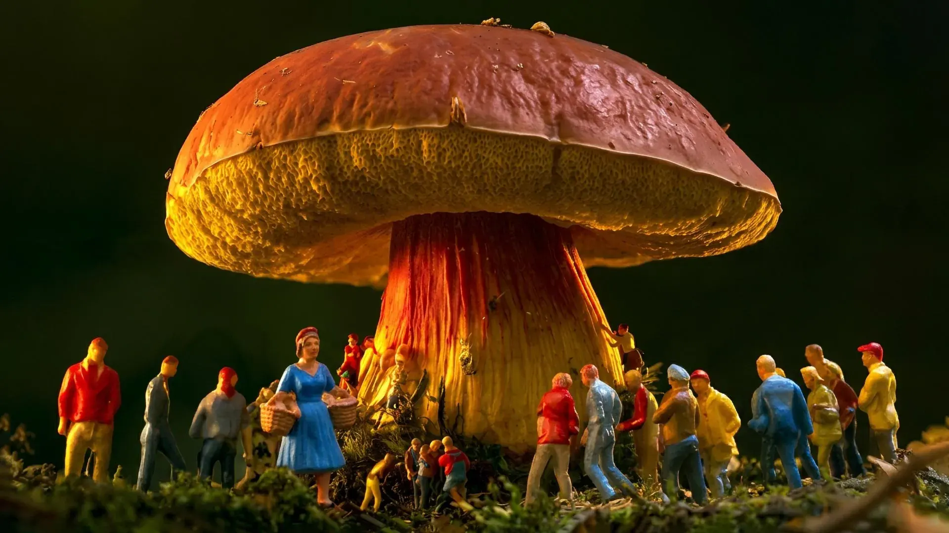
    People living thousands of miles apart have reported an extraordinarily strange experience after eating certain wild mushrooms. They begin seeing tiny human figures that appear to move through the real world, walking around objects, climbing over surfaces, and interacting with their surroundings.

    These visions are known as Lilliputian hallucinations, named after the miniature inhabitants of “Gulliver’s Travels,” who stand just 6 inches tall. For decades, researchers suspected that cultural beliefs and folklore might explain why people in different regions described such similar experiences.

    Now, a new study provides evidence that the phenomenon has a biological explanation.

    Researchers at the University of Utah (U) used DNA sequencing to identify Lanmaoa asiatica as the mushroom responsible for these unusual hallucinations in Southwest China and the northern Philippines. Surprisingly, their investigation found no evidence of any known psychoactive mushroom compounds, including psilocybin.

    The findings suggest that the mushroom may produce an entirely unfamiliar hallucinogenic substance. Identifying that chemical could help scientists better understand neurological disorders, unusual visual experiences, and the processes through which the brain creates our perception of reality.

    The research was conducted by Colin Domnauer, a doctoral student at the U, and Bryn Dentinger, a mycologist affiliated with the university and the Natural History Museum of Utah. Their findings were published in the journal Mycologia.

    In an interview with @theU, Domnauer explained how researchers traced the mushroom’s identity, what makes its effects so unusual, and why the discovery could have implications far beyond the study of fungi.

    A Century-Old Mystery of Tiny Human Hallucinations

    The scientific mystery dates back to 1934, when Western researchers learned that people in Papua New Guinea sometimes experienced vivid visions of miniature humans after eating wild mushrooms. The bizarre effect became known as “mushroom madness.”

    During the 1950s and 1960s, anthropologists and mushroom specialists traveled to Papua New Guinea to investigate. Their work pointed toward several possible species belonging to the Boletaceae (bolete) family.

    However, chemical testing failed to reveal any substances that could explain the hallucinations. Samples were even examined by Albert Hofmann, the scientist who first isolated psilocybin and discovered LSD. He, too, was unable to identify an active ingredient.

    With no chemical explanation available, some researchers began questioning whether the mushrooms were responsible at all. They suggested that the visions might instead arise from cultural expectations or traditional beliefs. By the end of the 1960s, scientific investigation of the phenomenon had largely stopped.

    The mystery resurfaced during the 1990s, when people in Yunnan, Southwest China, reported similar hallucinations after eating bolete mushrooms. Although the precise species remained uncertain, researchers suspected a recently described mushroom called Lanmaoa asiatica.

    Scientists Identify the Mushroom Behind the Strange Visions

    Another important clue appeared in 2024, when Domnauer learned of comparable experiences among an Indigenous community in the remote Northern Cordillera region of the Philippines.

    Community members described a wild edible mushroom called “Sedesdem” that could sometimes cause people to see miniature human figures. Similar mushrooms were known locally as “Nonda” in Papua New Guinea and “Jian shou qing” in Yunnan.

    These mushrooms have an important place in local food traditions, but consuming them without thoroughly cooking them can trigger the unusual hallucinations.

    The Philippine accounts represented a third independent report of wild mushrooms causing remarkably similar visions of tiny people.

    To investigate, Domnauer traveled to Yunnan and the Philippines, interviewed local residents, and collected specimens of L. asiatica along with other mushrooms belonging to the Lanmaoa genus.

    The fieldwork included the first scientific survey of fungi in the northern Philippines. Researchers then analyzed DNA from the collected mushrooms to establish their identities and evolutionary relationships.

    The results revealed that the mushrooms called Jian shou qing in China and Sedesdem in the Philippines were both L. asiatica.

    This was an unexpected finding because scientists had previously believed the species occurred only in China.

    Dentinger also visited Papua New Guinea in hopes of collecting specimens from the location where the mystery originally began. Unfortunately, unusually dry weather made it impossible to obtain samples for genetic comparison.

    Although the Papua New Guinea mushroom remains unverified, the evidence from China and the Philippines provides a compelling connection. Three geographically separated cultures have described the same highly specific hallucinations, and DNA testing has linked two of those cases to a single mushroom species.

    That consistency strongly suggests a shared chemical and neurological explanation rather than unrelated cultural influences.

    Hallucinations of Tiny Humans That Seem Remarkably Real

    What makes these hallucinations especially unusual is their striking similarity across different accounts.

    People typically describe seeing dozens or even hundreds of miniature figures, generally measuring between 3 and 30 centimeters tall. The figures appear with extraordinary clarity, often seeming as solid and convincing as actual people.

    According to Domnauer, approximately 90% of those experiencing the visions describe figures resembling elves, clowns, or other fairy-like characters, often wearing brightly colored clothing.

    Even more surprising is the way these imaginary figures seem to behave.

    Rather than floating through walls or ignoring their surroundings, they appear to follow familiar physical rules. They walk around obstacles, move across ordinary objects, and sometimes tumble from the edges of tables.

    Domnauer recalled one particularly unusual account involving someone eating soup. The person reported seeing tiny humans leap from a spoon into the bowl, where they began swimming. When the person took another spoonful, the miniature figures reportedly remained visible inside their mouth.

    Such detailed interactions distinguish these visions from many other forms of drug-induced hallucination.

    A Mysterious Psychedelic Compound Unlike Any Known Drug

    Scientists also investigated whether L. asiatica contains any of the chemicals already known to cause psychedelic experiences.

    Using both chemical testing and genetic analysis, the researchers searched for established psychoactive compounds found in other mushroom species.

    They found no trace of them.

    The absence of familiar hallucinogens was particularly intriguing because the mushroom’s effects differ so dramatically from those associated with known psychedelic drugs.

    Researchers now suspect that L. asiatica produces an unidentified psychoactive substance responsible for the tiny human hallucinations.

    The compound has not yet been isolated or chemically identified, so its exact nature and mechanism remain unknown.

    Discovering it could open an entirely new avenue of research into how certain chemicals alter visual perception.

    What the Mushroom Could Reveal About the Human Brain

    Although L. asiatica provides an unusually consistent trigger for Lilliputian hallucinations, the phenomenon is not limited to mushroom consumption.

    Stories involving miniature humanlike beings appear throughout folklore around the world. Similar hallucinations have also been documented in people experiencing alcohol withdrawal, dementia, macular degeneration, and other neurological conditions.

    These connections suggest that the visions may arise from a fundamental feature of the way the human brain processes visual information.

    However, scientists still do not fully understand the neurological mechanisms responsible for these experiences or how best to treat them when they occur in medical settings.

    This is where the mysterious mushroom could become especially valuable.

    Unlike the neurological and medical conditions in which Lilliputian hallucinations occur relatively rarely, L. asiatica can produce the phenomenon in a more consistent and recognizable way.

    If researchers can isolate the chemical responsible, they may gain a useful tool for investigating the brain circuits and processes that create these unusually detailed visual experiences.

    Such research could eventually improve scientific understanding of hallucinations associated with neurological diseases.

    The research team’s next major objective is to isolate and identify the suspected psychedelic compound.

    DNA Sequencing Reveals Four Previously Unknown Mushroom Species

    The investigation also produced important discoveries about fungal biodiversity.

    Rather than studying L. asiatica alone, Domnauer and Dentinger conducted the first comprehensive genomic analysis of the Lanmaoa genus.

    They examined 53 mushroom specimens collected from natural habitats and preserved in fungarium collections around the world.

    Genetic sequencing revealed 17 species within the group, including four species previously unknown to science.

    Remarkably, two of those newly identified species had already been preserved in existing collections in the United States. Their distinct identities had gone unrecognized until researchers examined their DNA.

    The analysis also indicated that L. asiatica is the only species in the genus currently known to produce psychoactive effects.

    Scientists are now working to reconstruct the group’s evolutionary history. They hope to determine how the unusual chemical trait developed and whether closely related mushrooms might produce comparable substances.

    The findings also demonstrate the scientific value of museum collections. Specimens preserved for years can contain biological discoveries that become apparent only when newer analytical techniques are applied.

    For Domnauer, the hidden diversity found in these collections is a reminder that many species and potentially important biological characteristics remain undiscovered, even among organisms that scientists have already collected.

    Exploring Fungi Could Lead to New Medicines and Discoveries

    The broader implications extend well beyond unusual hallucinations.

    Fungi remain one of the least thoroughly explored groups of organisms, and researchers believe their chemical diversity could provide valuable clues about biology, medicine, and the nervous system.

    One of the most famous examples is penicillin, the antibiotic originally obtained from a fungus that transformed the treatment of bacterial infections and became one of the most consequential medical discoveries of the last century.

    Scientists hope continued exploration of fungal biodiversity could reveal other medically useful compounds and previously unknown biological processes.

    The mysterious effects of L. asiatica illustrate just how much remains to be learned about the fungal kingdom. A mushroom long consumed by communities in Asia may contain a substance capable of revealing new information about the human brain and how it creates conscious experience.

    The research received funding from the National Science Foundation (award #DEB-2114785).


    Originally published by www.sciencedaily.com.

    Read the full article here.

  • Scientists propose a way to detect dangerous “mirror life” before it exists

    Scientists propose a way to detect dangerous “mirror life” before it exists

    Scientists have proposed what they believe is the first way to detect “mirror life,” a hypothetical form of life built from molecules that are reversed versions of those found in living organisms on Earth.

    Researchers from the University of Strathclyde and Durham University have developed a proposed detection technique that uses molecules capable of emitting light with distinct left and right-handed properties. By analyzing these light signals, scientists may eventually be able to distinguish conventional organisms from their mirror-image counterparts.

    Although mirror life does not currently exist, some experts consider its potential creation a serious biological safety concern. These artificial organisms could behave in unpredictable ways when interacting with Earth’s ecosystems and might evade certain natural defenses that protect people, animals, and plants.

    The likelihood of mirror life emerging is considered low, but its possible consequences could be severe enough to justify developing detection methods in advance.

    Why Mirror Life Could Pose a Biological Threat

    Concerns about mirror life have prompted scientists to recommend restrictions on research intended to produce organisms capable of reproducing using mirror-image biological molecules.

    The potential dangers have also attracted attention from governments and international organizations, including the UK Government Office for Science and the United Nations Secretary General’s Scientific Advisory Board.

    The underlying science involves a property of molecules known as chirality, often described as molecular “handedness.” Just as a person’s left and right hands are mirror images that cannot be perfectly aligned, some molecules can exist in two forms that have identical chemical components but different three-dimensional arrangements.

    These subtle structural differences can have major biological consequences. Two mirror-image versions of a molecule contain the same atoms connected in the same order, yet the human body may respond to them very differently. For example, one form of a medication could successfully treat a disease, while its mirror-image counterpart might be ineffective or even harmful.

    Scientists are concerned that organisms assembled from reversed biological building blocks could interact with existing life in unexpected ways.

    Importantly, the researchers emphasize that mirror life remains entirely hypothetical. Their proposed detection method is intended as a precaution, not a response to an existing biological threat.

    How Light Could Reveal Mirror-Image Life

    The proposed approach relies on a phenomenon called circularly polarized luminescence (CPL), which can reveal the molecular handedness, or chirality, of certain substances and biological structures.

    Light can exhibit different polarization patterns, including forms that rotate in opposite directions. By studying these patterns, researchers can obtain information about the three-dimensional arrangement of molecules.

    The underlying concept has a surprisingly familiar application: the polarized light technology used in modern 3D movie glasses to create the illusion of depth.

    The research, published in Royal Society Open Science under the title ‘Circularly Polarized Luminescence (CPL) as a Contingency Assay for “Mirror Life” Bio-Hazards’, was led by Dr. Lewis MacKenzie of the University of Strathclyde’s Department of Pure and Applied Chemistry, working with Professor Robert Pal of Durham University.

    Dr. MacKenzie said: “We don’t expect mirror life to appear tomorrow, but if it did arise, the consequences could be severe, and it makes sense to develop detection methods before they are needed. Professor Pal and I had been working on CPL technologies before recent concerns became prominent, and guarding against mirror life was a clear fit for our research.”

    A Molecular Fingerprint for Detecting Mirror Life

    To identify a possible mirror organism, the researchers suggest using specialized fluorescent probes that attach to particular structures inside cells.

    Once bound to their targets, these probes would generate characteristic CPL signals. Scientists could analyze the resulting patterns to determine whether a biological sample contains the molecular handedness associated with ordinary terrestrial life or its reversed equivalent.

    In effect, the light signals could serve as a molecular fingerprint, revealing differences that might otherwise be difficult to identify.

    However, relying on a single fluorescent probe could produce misleading results. To improve reliability and reduce the risk of false readings, the team proposes combining several probes designed to recognize different cellular structures.

    By examining multiple signals together, researchers hope to develop a more dependable way to distinguish conventional organisms from hypothetical mirror life.

    Testing the Technology Without Creating Mirror Organisms

    One important advantage of the proposed technique is that scientists could begin investigating and refining it using ordinary biological samples. There would be no need to manufacture mirror organisms simply to explore whether the underlying detection principles work.

    The researchers also believe the method could eventually be incorporated into several types of scientific equipment, potentially allowing mirror-life screening in different environments.

    Professor Pal has already demonstrated laboratory-scale CPL detection technologies involving microscopes, cameras, and biological cell plate readers, which are instruments commonly used to examine large numbers of cell samples.

    However, the complete detection method specifically designed to identify mirror life has not yet been experimentally demonstrated. Further research will be necessary to determine whether the proposed approach can reliably distinguish between different forms of molecular handedness in biological samples.

    Professor Pal added: “CPL detection relies on the same fundamental concept that allows modern 3D cinema glasses to create a stereoscopic image.

    “This CPL detection approach can be adapted to many types of systems for analysis of cells, including biological plate readers, microscopes, and cameras. This means our mirror life assay approach could be generalized to many environments depending on end user needs.”


    Originally published by www.sciencedaily.com.

    Read the full article here.

  • These tiny snakes roll four times faster than they slither

    These tiny snakes roll four times faster than they slither

    Tiny snakes found in Southeast Asia have developed an extraordinary way to escape danger. Instead of relying on the familiar side-to-side motion of slithering, dwarf reedsnakes can rapidly roll across the ground, reaching speeds about four times faster than their usual movement.

    Scientists have now uncovered how these small reptiles accomplish such an unusual feat. By studying video recordings captured from multiple angles, researchers discovered that the snakes coordinate their movements with remarkable precision. While the rear portions of their bodies rotate rapidly, their heads remain relatively steady, facing forward and staying at a nearly constant height.

    [See link to video after the end of the article.]

    The findings reveal how an animal can achieve an unexpected form of movement through specialized behavior and body control, even without the physical structures normally needed for rolling.

    Why Rolling Is So Difficult for Animals

    Rolling might seem like an effective way to travel quickly, but it presents a major challenge for living creatures.

    A wheel can spin continuously around a fixed axle, allowing it to rotate without disturbing whatever it supports. Animals, however, have no comparable mechanism separating their rotating bodies from their heads.

    If an animal’s entire body spins together, its head and sensory organs must also rotate. This can make it difficult to maintain balance, adjust body position, and steer in the intended direction. As rolling speed increases, these problems become even harder to manage.

    Yet dwarf reedsnakes belonging to the genus Pseudorabdion have found a way around these limitations.

    These small Southeast Asian snakes are among the few animals known to deliberately roll as a means of escaping potential predators. Their unusual behavior has appeared in field guides and was previously documented during a field observation, but scientists had not fully understood the movements that made it possible.

    Tiny Snakes Roll Four Times Faster Than They Slither

    To investigate this remarkable escape strategy, researchers filmed dwarf reedsnakes from several angles and carefully examined how different parts of their bodies moved during rolling.

    The results revealed impressive speeds. While rolling, the snakes reached peak forward velocities ranging from 1.72 to 3.69 meters per second, approximately four times their maximum speed when moving through lateral undulation, the familiar side-to-side motion used by most snakes.

    Despite traveling so quickly, the snakes were able to follow a narrow racetrack measuring just 20 centimeters across, demonstrating that their rolling movements are not simply uncontrolled tumbling.

    Closer examination of the recordings revealed a surprisingly intricate sequence of movements.

    Rather than spinning their entire bodies together, the snakes alternate between rolling on one side and swinging a curved section of their bodies downward beside their heads. The researchers called this downward sweeping movement a “stroke.”

    During each successive cycle, the side touching the ground switches, and the stroke changes direction from left to right or right to left. By repeating this carefully timed pattern, the snakes generate continuous forward rolling motion.

    The Remarkable Secret Behind Their High-Speed Escape

    One of the most surprising discoveries involved what happens to the snakes’ heads during these rapid movements.

    Although the rear portions of their bodies undergo substantial rotation, the snakes keep their heads pointed in the direction they are traveling. Rather than spinning along with the rest of the body, the head remains comparatively stable.

    The researchers also found that the snakes maintain a relatively consistent head height above the ground. This stabilization becomes especially noticeable at higher speeds, when the snakes tend to hold their heads elevated and minimize upward and downward movements.

    Keeping the head steady while the rest of the body rotates may help the animals maintain control and navigate effectively during their rapid escapes.

    This ability is particularly remarkable because the snakes accomplish it without a mechanical separation between the rotating and stabilized portions of their bodies.

    What Rolling Snakes Reveal About Evolution

    The discovery demonstrates how sophisticated coordination can allow animals to overcome physical limitations that might otherwise make certain types of movement impractical.

    Although dwarf reedsnakes lack anything resembling a wheel’s axle, they effectively divide their bodies into regions with different functions. Much of the body performs the powerful rotational movements needed for propulsion, while the head remains stabilized to support controlled travel.

    This unusual strategy allows the snakes to combine speed, directional control, and rapid body rotation in a way that would be difficult to predict from their anatomy alone.

    More broadly, the findings highlight an important principle of animal evolution: physical structure does not necessarily determine the full range of movements an animal can perform. Innovative behaviors and precise muscular coordination can open up entirely new possibilities, allowing creatures to accomplish feats that their body shapes might initially suggest are impossible.


    Originally published by www.sciencedaily.com.

    Read the full article here.

  • This 150-million-year-old bird had dinosaur teeth and a strange secret

    This 150-million-year-old bird had dinosaur teeth and a strange secret

    A remarkably preserved 150-million-year-old bird fossil discovered in China is challenging long-standing ideas about how birds evolved. Rather than developing increasingly sophisticated flight abilities, some of the earliest birds appear to have taken a very different evolutionary path, including losing the ability to fly altogether.

    The newly identified species, Yuezhuornis yangyuanensis, lived during the Late Jurassic period (150-148 million years ago). Described in a study published in Nature, the crow-sized creature provides evidence that early bird evolution was much more complex than scientists once believed. Instead of evolving primarily to become better flyers, ancient birds were adapting to a variety of environments and ways of life, including those that favored living on the ground.

    Researchers from the Institute of Vertebrate Paleontology and Paleoanthropology (IVPP) of the Chinese Academy of Sciences (CAS) and the Fujian Institute of Geological Sciences uncovered the fossil in the recently recognized Zhenghe Fauna of Fujian Province in southeastern China. According to their findings, Yuezhuornis represents the earliest known example of a bird lineage losing its ability to fly.

    An Ancient Bird With Dinosaur Teeth and Claws

    One of the most remarkable features of Yuezhuornis is its unusual combination of bird and dinosaur characteristics. Although it possessed skeletal features associated with birds, it also retained anatomical traits that would have been familiar among small predatory dinosaurs.

    Its jaws contained closely spaced, serrated teeth resembling those of deinonychosaurs, a group of meat-eating dinosaurs that included agile predators related to birds. Such teeth are not normally associated with early birds.

    Even more striking was its second toe, which could bend backward unusually far and carried a curved, sickle-shaped claw. This specialized feature is strongly associated with deinonychosaurs, including dromaeosaurids and troodontids, two groups of predatory dinosaurs.

    “When we examined Yuezhuornis, we were struck by its extraordinary mix of features,” said Dr. Min Wang, corresponding author of the study. “It possesses bird-like pelvic and hindlimb structures yet retains the specialized predatory foot and teeth of a small meat-eating dinosaur. It shows us that early bird evolution was far more complicated than we previously realized.”

    Where Does This Strange Jurassic Bird Fit in Evolution?

    To understand how Yuezhuornis was related to other prehistoric birds and dinosaurs, scientists carried out detailed analyses of its evolutionary relationships. They used two approaches, maximum parsimony and Bayesian tip-dating, to compare anatomical characteristics and reconstruct the species’ position on the evolutionary tree.

    Maximum parsimony identifies evolutionary relationships that require the fewest changes in characteristics, while Bayesian tip-dating also incorporates statistical estimates of evolutionary history and information about when fossils lived.

    Both methods produced consistent results, placing Yuezhuornis within Avialae, the evolutionary group that includes modern birds and their closest extinct relatives.

    “The results consistently placed Yuezhuornis firmly within Avialae (the group containing modern birds and their closest extinct relatives), just crownward of Archaeopteryx and as a sister taxon to Zhengheornis,” said Dr. Zhonghe Zhou, a co-author of the study.

    This placement is particularly important because Archaeopteryx, which also lived roughly 150 million years ago, is among the most famous early bird relatives. The findings indicate that Yuezhuornis belonged to an early branch of bird evolution rather than simply being a small predatory dinosaur with bird-like characteristics.

    Evidence That Some Early Birds Lost the Ability to Fly

    The researchers also examined how Yuezhuornis might have lived. Using measurements of its skeletal proportions and statistical comparisons with other animals, they investigated whether its anatomy was better suited for flying or moving along the ground.

    The results showed that its limb proportions closely resembled those of modern ground-dwelling birds and certain other prehistoric dinosaurs closely related to birds. This similarity suggests that Yuezhuornis spent its life on the ground and was likely unable to fly.

    The findings point to an unexpected development in the history of birds. Rather than simply progressing toward stronger wings and more efficient flight, some early bird lineages appear to have moved in the opposite direction, adapting to lifestyles in which flying was no longer necessary.

    This challenges the traditional assumption that the earliest stages of bird evolution were driven primarily by the demands of flight. Instead, birds were already exploring different ecological roles and developing a surprisingly wide range of body forms.

    A 150-Million-Year-Old Discovery Rewrites Bird Evolution

    The discovery of Yuezhuornis suggests that the evolution of birds was never a simple, one-directional process. Even during the Late Jurassic, when the earliest bird lineages were emerging, some species were developing adaptations that favored life on land rather than in the air.

    Its unusual combination of bird-like anatomy, dinosaur-style predatory features, and apparent flightlessness provides a revealing glimpse into that evolutionary experimentation. The fossil shows that early birds were more anatomically and ecologically diverse than previously recognized.

    It also underscores the scientific importance of the Zhenghe Fauna, a fossil assemblage that is helping researchers investigate a poorly understood period in bird evolution. As additional discoveries emerge from this region of China, scientists may gain an even clearer picture of the surprising evolutionary paths taken by the earliest birds.


    Originally published by www.sciencedaily.com.

    Read the full article here.

  • Birds are more afraid of women than men. Scientists have no idea why

    Birds are more afraid of women than men. Scientists have no idea why

    Birds living in cities appear to react differently to men and women, and scientists are struggling to explain why. An international research team discovered that urban birds, including great tits, house sparrows, and blackbirds, tend to fly away sooner when approached by women than by men. The surprising behavior raises new questions about how birds perceive the people around them.

    The study took place in five European countries, where male and female participants (matched for height and clothing) walked directly toward birds in city parks and other green spaces.

    On average, men were able to approach about one meter closer than women before the birds flew away. The researchers measured this response by recording how close a person could get before a bird took flight (termed flight initiation distance).

    Published in the British Ecological Society journal People and Nature, the findings showed the same pattern in Czechia, France, Germany, Poland, and Spain. The difference also appeared across all 37 bird species examined, including easily startled magpies and pigeons that typically allow people to approach much more closely.

    Why Are Birds More Afraid of Women?

    The researchers believe their findings indicate that city birds can distinguish between male and female humans. However, they have yet to determine which characteristics the birds recognize or why they appear more cautious around women.

    Professor Daniel Blumstein of the University of California, Los Angeles, who helped conduct the research, said the results were convincing despite the lack of an explanation.

    “I fully believe our results, that urban birds react differently based on the sex of the person approaching them, but I can’t explain them right now. We used bleeding-edge comparative analysis techniques that showed our findings were consistent across cities and species, but we simply don’t have a conclusive explanation yet.”

    Dr. Yanina Benedetti of the Czech University of Life Sciences Prague, another member of the research team, said the discovery challenged her expectations about how birds respond to human observers.

    “As a woman in the field, I was surprised that birds reacted to us differently. This study highlights how animals in cities ‘see’ humans, which has implications for urban ecology and equality in science. Many behavioral studies assume that a human observer is neutral, but this wasn’t the case for urban birds in our study.”

    What Are Birds Detecting in Humans?

    Scientists have proposed several possible explanations for the difference. Birds might be responding to chemical signals such as pheromones, variations in body shape, or differences in how men and women walk. For now, however, these ideas remain speculative, and additional experiments will be needed to identify the actual cause.

    Dr. Federico Morelli of the University of Turin, a co-author of the study, said the unanswered question is one of the discovery’s most fascinating aspects.

    “This is maybe the most interesting part of our study,” said Dr. Federico Morelli from the University of Turin, a co-author of the study. “We have identified a phenomenon, but we really don’t know why. However, what our results do highlight is the birds’ sophisticated ability to evaluate their environment.”

    The findings suggest that urban birds may be sensitive to human characteristics that people themselves rarely notice. Identifying those signals could help researchers better understand how wildlife assesses potential threats in environments shared with humans.

    Dr. Benedetti said: “Urban birds clearly react to subtle cues that humans do not easily notice. Follow-up studies could focus on individual factors such as movement patterns, scent cues, or physical traits, testing them separately rather than grouping them under observer sex. This approach would help identify the specific cues birds detect.”

    More Research Is Needed to Explain the Bird Behavior

    Although the investigation included 2,701 observations, the researchers emphasize that their findings are still preliminary. Further studies will be necessary to establish whether the difference between birds’ responses to men and women consistently appears in other settings and populations.

    For now, the discovery points to an intriguing possibility: Birds sharing our cities may be paying attention to subtle differences between people that scientists have yet to fully understand.


    Originally published by www.sciencedaily.com.

    Read the full article here.

  • Oxford scientists discover how viruses build themselves without a blueprint

    Oxford scientists discover how viruses build themselves without a blueprint

    Researchers at the University of Oxford have observed virus-like particles assembling themselves one molecule at a time, uncovering the surprisingly simple rules that allow complex biological structures to form with remarkable reliability. The findings, published in Nature, offer a detailed look at a process that scientists have struggled to observe directly for decades.

    Understanding how viruses construct their protective protein shells, and how that construction might be interrupted, could lead to new antiviral drugs, improved vaccines, and other medical treatments. The discovery helps explain how individual proteins consistently organize themselves into the correct structures despite having thousands of possible ways to connect.

    The researchers found that viral assembly resembles navigating a complicated maze, where only certain paths lead to lasting progress. At first, protein components form weak connections that can easily break apart if the arrangement is unsuccessful. This flexibility allows the molecules to keep trying different combinations until they form particular closed structures. Once multiple connections hold these structures together, they become far more stable.

    These stable arrangements serve as molecular checkpoints, narrowing the enormous number of possible assembly routes into a much smaller set of successful pathways. One especially important milestone occurs when five larger protein building blocks connect to form a closed pentagonal ring. This is the first highly stable structure in the assembly process. Once it forms, each subsequent stable stage requires fewer additional building blocks, allowing the particle to grow more quickly.

    Co-lead author Dr. Roi Asor (Department of Chemistry and Kavli Institute for Nanoscience Discovery, University of Oxford) explained: “A virus has to solve an extraordinary construction problem. Its components somehow have to find the right arrangement among a huge number of possibilities, without a blueprint or machinery directing the process. We can now watch what happens molecule by molecule and see the physical rules that make it possible.”

    Watching Virus Particles Assemble Molecule by Molecule

    Viruses store and protect their genetic material within organized protein shells known as capsids. Depending on the virus, these shells may consist of tens or even thousands of individual protein components. Remarkably, in many cases, the proteins can arrange themselves into the correct structure without any external machinery directing their assembly.

    Although scientists have investigated viral self-assembly for decades, capturing the process as it unfolds has been a major challenge. Many of the intermediate structures exist only briefly and appear in very small quantities. As a result, traditional methods have generally allowed researchers to examine the starting materials and finished particles without clearly showing how one transforms into the other.

    To overcome this limitation, the Oxford researchers studied an engineered virus-like particle composed of 60 protein units. They used mass photometry, a technique developed at Oxford that determines the mass of individual molecules by measuring the light they scatter. By combining this technology with a new approach for keeping individual molecules confined and continuously observable, the team could repeatedly measure the mass of a growing particle as additional proteins attached.

    This approach allowed the scientists to follow the construction process as it happened, effectively tracking the addition of individual molecular building blocks over time.

    Co-first author PhD student Dan Loewenthal (Department of Chemistry and Kavli Institute for Nanoscience Discovery, University of Oxford) said: “This method lets us study the assembly process directly, we just take a video!”

    Co-lead author Professor Philipp Kukura (Department of Chemistry and Kavli Institute for Nanoscience Discovery, Oxford University) said: “Until now, much of our understanding of how these structures assemble has had to be reconstructed from snapshots or theoretical models. Being able to both quantify the underlying interactions and follow one particle as it grows changes that. We can see the important intermediate structures appear and deduce a detailed model of the construction process.”

    The Surprising Trick That Makes Viral Assembly Work

    The results help explain how a process involving an overwhelming number of possible molecular arrangements can consistently produce the correct structure. Rather than exploring every possible route, the proteins progress through a relatively small number of stable intermediate stages that guide assembly toward completion.

    The findings also clarify why the earliest stage of construction, called nucleation, is particularly difficult and why growth becomes substantially faster after the first stable structure appears.

    A key part of this efficiency comes from the ability of protein building blocks to make mistakes without permanently disrupting the process. Weak initial connections allow incorrect combinations to separate, giving the proteins another opportunity to find a better arrangement. When enough components eventually connect in the correct closed configuration, the resulting structure becomes stable enough to support further growth.

    Dr. Asor added: “The weak interactions give the system room to make mistakes. Most encounters don’t have to be successful: the components can separate and try again. But once enough of them come together in the right closed arrangement, the structure becomes stable, and assembly can move forward. That combination of trial and error followed by locking in successful structures is what makes the process so reliable.”

    New Possibilities for Antiviral Drugs and Vaccines

    By identifying the molecular interactions that control viral assembly, scientists may eventually be able to develop treatments that interfere with the construction of viral protein shells. The same insights could also support efforts to engineer virus-like particles for vaccines and other therapeutic applications.

    Co-author Dr. Jack Tan (MRC Weatherall Institute of Molecular Medicine and CAMS-Oxford Institute) noted: “The ability to understand these processes at the molecular level could have important applications in vaccine development and antivirals that disrupt viral assembly.”

    The implications may extend far beyond viruses. Self-assembly is essential throughout biology, helping create everything from the internal skeletons of cells to specialized molecular compartments and large protein complexes.

    By making it possible to observe these intricate construction processes one molecule at a time, the Oxford team’s approach offers researchers a powerful new way to investigate how complex biological structures emerge from relatively simple interactions.


    Originally published by www.sciencedaily.com.

    Read the full article here.